Ear & Balance Disorders
Cholesteatoma
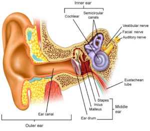
 A cholesteatoma is an abnormal skin growth in the middle ear behind the eardrum. Although a cholesteatoma is not a tumor, it can increase in size and destroy the surrounding delicate bones of the middle ear leading to hearing loss, drainage from the ear, dizziness, and other complications due to injury to the surrounding structures. Repeated infections, a hole in the ear drum, or pulling inward of the eardrum from eustachian tube dysfunction can allow skin into the middle ear and form a cholesteatoma. The eustachian tubes convey air from the back of the nose into the middle ear to equalize ear pressure (ear popping when on a plane or with changes with altitude.)
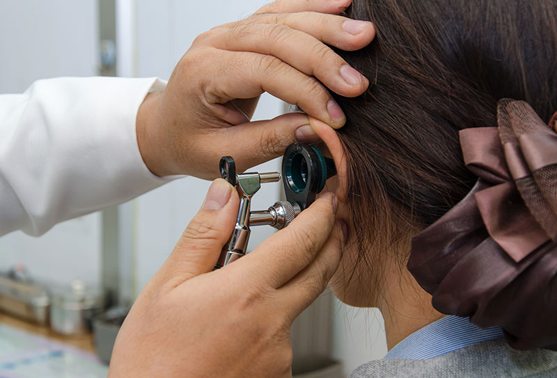
Individuals with chronic or recurrent drainage from the ears, chronic ear infections, ear pain, hearing loss or dizziness will require an examination by an otolaryngologist. Our doctors at C/V ENT Surgical Group will perform a thorough ear exam and evaluation to determine the presence of a cholesteatoma. Initial treatment may consist of a careful ear cleaning, antibiotics, and ear drops. Initial treatment will aim to stop the drainage. A hearing test and a CT scan maybe performed to determine the hearing level in the ear and the extent of destruction caused by the cholesteatoma. Definitive therapy will usually require surgery. Cholesteatoma is a serious but treatable ear condition, which can be diagnosed only by medical examination. Bone erosion can cause the infection to spread into the surrounding areas, including the inner ear and brain. If untreated, deafness, brain abscess, meningitis, and, rarely, death can occur.
Dizziness and Vertigo
Feeling unsteady or dizzy can be caused by a variety of factors such as poor circulation, inner ear disease, medications, trauma, infection, allergies, and/or neurological disease.

The sensation of vertigo is usually due to an issue with the inner ear. A vertigo attack may cause sudden nausea, vomiting and heavy sweating. Severe vertigo causes a loss of balance and can cause you to fall. During vertigo, small head movements and changes in body position will often make the symptoms worse. An episode of vertigo may last seconds, minutes or hours. Once you are over the first episode, it may never return. However, sometimes symptoms may recur off and on over several weeks or longer, depending on the cause. There may be ringing in the ears or hearing loss, which may be temporary or permanent.
Common causes of vertigo include: Benign Positional Vertigo, Meniere’s disease, migraine, infection, injury, and allergy.
Our experts at C/V ENT Surgical Associated will obtain a thorough history and examination and guide the treatment of your vertigo. A hearing test is often required to evaluate the function of the ear. Treatment will depend on the likely cause of your dizziness.
Middle Ear Infection

Hearing Loss
There are 2 types of hearing loss: conductive and sensorineural. One or both kinds can occur.
A conductive hearing loss occurs when sound waves do not reach the inner ear. Sound waves may be disrupted before they reach the inner ear due to a variety of conditions. The ear canal can be blocked by wax, infection, a tumor, or a foreign object. The eardrum can be injured or infected. Abnormal bone growth, infection, or tumors in the middle ear can block sound waves.
A sensorineural hearing loss occurs when sound waves are not processed correctly by the inner ear, the 8th cranial nerve, or central nervous system.
Our specialists at C/V ENT Surgical Group will perform a thorough examination to determine the type and cause of your hearing loss and discuss treatment accordingly.
Hyperacusis
Individuals with hyperacusis have difficulty tolerating sounds which do not seem loud to others. Hyperacusis is a condition that arises from a problem in the way the brain’s central auditory processing center perceives noise. It can often lead to pain and discomfort.
Many people experience sensitivity to sound, but true hyperacusis is rare, affecting approximately one in 50,000 individuals. The disorder can affect people of all ages in one or both ears. Individuals are usually not born with hyperacusis, but may develop a narrow tolerance to sound.
Individuals who suspect they may have hyperacusis should seek an evaluation by one of experts. There are no specific corrective surgical or medical treatments for hyperacusis. However, sound therapy may be used to “retrain” the auditory processing center of the brain to accept everyday sounds.
Perforated Eardrum
A hole or rupture in the eardrum, a thin membrane that separates the ear canal and the middle ear, is called a perforated eardrum. A perforated eardrum is often accompanied by decreased hearing and sometimes liquid discharge. The perforation may be accompanied by pain, if it is caused by an injury or becomes infected. A perforation can occur from injury, infection, or chronic Eustachian tube disorders.
Traumatic causes include direct trauma such as with use of a bobby pin or Q-tip, skull fracture, or slap to the ear. Middle ear infections may lead to spontaneous rupture of the eardrum with infected or bloody drainage from the ear. In patients with chronic Eustachian tube problems, the ear drum may become progressively weakened and a perforation to occur. On some occasions, a small hole may remain in the eardrum after a previously placed pressure-equalizing tube falls out or is removed.
Most eardrum holes resulting from injury or an acute ear infection heal on their own. The chance of a perforation healing on its own depends on the size of the perforation. If the perforation does not spontaneously heal, then surgical options can be considered.
Our specialists at C/V ENT Surgical Group will advise you regarding the proper care of a hole in the eardrum.
Swimmer’s Ear/Otitis Externa
Swimmer’s ear or acute otitis externa is a painful condition resulting from inflammation, irritation, or infection of the outer ear canal. These symptoms often occur after water gets trapped in your ear, with subsequent spread of bacteria or fungal organisms. When water is trapped in the ear canal, bacteria that normally inhabit the skin and ear canal multiply, causing infection of the ear canal.
Other causes of otitis externa include:
- Contact with polluted water
- Excessive cleaning of the ear canal
- Damage to the skin of the ear canal following water irrigation to remove wax
- A cut in the skin of the ear canal
- Other skin conditions affecting the ear canal, such as eczema
Our ENT surgeons at C/V ENT Surgical Group will examine your ears, carefully clean your ear canal and typically prescribe eardrops that inhibit bacterial or fungal growth and reduce inflammation. If the ear canal is swollen shut, a wick may be placed in the canal so the antibiotic drops will enter the swollen canal more effectively. Follow-up appointments are very important to monitor improvement or worsening, to clean the ear again, and to replace the ear wick as needed. At C/V ENT Surgical Group, we have specialized equipment and expertise to effectively clean the ear canal and treat swimmer’s ear.
Tinnitus
Tinnitus is the perception of sound without an external source being present. The noise might be a ringing, clicking, hiss, or roar. It can vary in pitch and may be soft or quite loud. For some people, tinnitus is a minor nuisance. But for others, the noise can make it hard to hear, work, and even sleep. About one in five people with tinnitus have bothersome tinnitus, which distresses them and negatively affects their quality of life. Tinnitus may be an intermittent or continuous sound in one or both ears. Prior to any treatment, it is important to undergo a thorough examination and evaluation by our specialists at C/V ENT Surgical Group as an essential part of the treatment will be your understanding of tinnitus and its causes.
Tinnitus may be caused by different parts of the hearing system. In the outer ear, excessive ear wax can result in tinnitus. Middle ear problems including middle ear infections may lead to tinnitus. Most subjective tinnitus associated with the hearing system originates in the inner ear. Damage and loss of the tiny sensory hair cells in the inner ear may be commonly associated with the presence of tinnitus. In certain cases, tinnitus may develop with loud noise exposure even before hearing loss. Medications can also damage inner ear hair cells and cause tinnitus. As we age, the incidence of tinnitus increases.
Tinnitus may also originate from lesions on or in the vicinity of the hearing portion of the brain. These include a variety of uncommon disorders including vestibular schwannoma (acoustic neuroma) and damage from head trauma.
Another category is pulsatile tinnitus that sounds like one’s heartbeat or pulse. Infrequently, pulsatile tinnitus may signal the presence of cardiovascular disease or a vascular tumor.
